A red swelling in the inner corner of the eye is usually the tear gland of the third eyelid, slipped out of place. We tuck it back in rather than remove it, so it goes on making tears.
Plate VICherry eye: the gland of the third eyelid, out and backProlapsus glandulae membranae nictitantis
Third eyelidmembrana nictitansThird eyelidmembrana nictitansIts dark leading edgeIts dark leading edgeGland of the third eyelidout of placeGland of the third eyelidout of placeThe gland, back in placehidden behind the eyelidThe gland, back in placehidden behind the eyelidClear front of the eyecorneaClear front of the eyecorneaT-shaped cartilagein sectionT-shaped cartilagein sectionThe glandflipped over the edgeThe glandflipped over the edgeThe glandback at its baseThe glandback at its basePocket stitchdissolvingPocket stitchdissolving
The gland of the third eyelid has slipped out over the eyelid’s edge and swollen. Fig. 2, in the circle, shows it in section, flipped out of place.
1. Third eyelid membrana nictitans
2. Its dark leading edge
3. Gland of the third eyelid, out of place
3. Gland back in place, hidden behind the third eyelid
4. Clear front of the eye cornea
5. T-shaped cartilage, in section (Fig. 2)
6. The gland, flipped over the edge (Fig. 2)
6. The gland, back at the base (Fig. 2)
7. Pocket closed with a dissolving stitch (Fig. 2)
Tap a number on the plate or in the list to see its label.
Most often in
Young dogs
Anaesthetic
General
Collar
Around 10 to 14 days
Stitches
Dissolving
Your vet: Dr John Alexander MRCVS, 20 years in practice. RCVS Register
A written estimate before anything goes ahead
Aftercare included: a written plan, check-ups, and help day and night
The 30-second version
Cherry eye is the tear gland of the third eyelid popping out of place. It shows as a smooth red swelling in the inner corner of the eye.
It is rarely an emergency, but it does need fixing. The longer the gland is out, the more swollen and irritated it becomes.
We put the gland back, usually with the pocket technique. We don’t remove it as a first choice, because it makes a large share of the eye’s tears.
Recovery means a buster collar, eye drops and a check at around 10 to 14 days.
Dogs have a third eyelid, a thin fold of pink tissue in the inner corner of each eye that sweeps across to protect it. It is stiffened by a small T-shaped piece of cartilage, and at its base sits a tear gland. That gland makes a large part of the watery tears that keep the eye healthy: around a third or more.
The gland is normally held down by a small band of tissue. In some dogs that hold is weak, and the gland flips up over the edge of the third eyelid. Out in the air it swells and turns red, which is why it looks like a cherry.
It is most common in young dogs, often before their second birthday, and in breeds such as English and French Bulldogs, Cocker Spaniels, Beagles, Shih Tzus, Lhasa Apsos, Boston Terriers, Cane Corsos and Neapolitan Mastiffs. It can affect one eye or both, and the second eye sometimes follows weeks or months later. Cats get it rarely; Burmese and Persians most often.
Signs to look out for
A smooth, red or pink swelling in the inner corner of the eye
A swelling that comes and goes at first, then stays out
A watery or sticky eye, and tear staining below it
Rubbing or pawing at the eye
A red, sore-looking eye
A painful eye is an emergency
Cherry eye itself is rarely urgent, but squinting, a cloudy eye or a sudden change in the eye is. Call us the same day on 01843 808344.
How we find out
We examine both eyes closely and confirm that the swelling is the gland, and not something that can look similar, such as a bent (scrolled) cartilage in the third eyelid or a growth.
A paper strip test measures how many tears each eye makes, so we know the gland’s starting point. A drop of orange dye checks the surface of the eye for ulcers. We look at the other eye too.
The operation
Why we put the gland back
In the past the gland was often cut away. We now know that removing it raises the risk of dry eye later in life, a condition called keratoconjunctivitis sicca that needs drops every day for good. So the aim is always to put the gland back and keep it working.
The pocket technique
Under general anaesthetic, two small cuts are made in the lining on the inner side of the third eyelid, the side that faces the eye, one above the gland and one below. The gland is tucked into the pocket between them, and the cuts are closed over it with a very fine dissolving stitch, tied so that it doesn’t rub on the eye. The diagram shows the change, and Fig. 2 in its circle shows the third eyelid in section.
Large or long-standing glands sometimes need a different way of anchoring them. John will explain which suits your dog.
In published studies the pocket technique works first time in most dogs, but a small number of glands slip out again and need a second operation.
Recovery, week by week
Every animal heals at its own pace, and we’ll give you a written plan for yours. As a guide:
Straight away
A buster collar at all times, so your dog can’t rub the eye. Eye drops or ointment and pain relief as prescribed.
The first few days
The third eyelid looks swollen and there may be a little discharge. It settles over a week or so.
Around 10 to 14 days
A check that the gland is staying put and the eye is comfortable. The stitches dissolve on their own.
Long term
Keep an eye on the other eye as well, and let us know straight away if a swelling comes back.
Every anaesthetic and operation carries some risk, and we will go through yours before anything is booked. With cherry eye the main things we watch for are the gland slipping out again, swelling, and irritation from the stitch. A gland that has been out for a long time may already be making fewer tears, which is why we measure tear production before and after.
Your questions answered
Will it go back on its own?
Occasionally it slips back for a while, but it usually comes out again. Pushing it back by hand is not a lasting fix, and the longer it is out, the more swollen it becomes.
Why not just remove it?
The gland makes a large share of the eye’s tears. Removing it raises the risk of dry eye, which needs drops for life. Putting it back keeps it working.
Will the other eye get it?
It can. Many dogs only ever have one, but in some the second eye follows, so we check both.
Will insurance cover it?
It depends on your policy, and some insurers treat it as a breed-related condition. Check your policy wording before any planned procedure.
What it costs
Every operation is different, so we give you a written estimate once John has examined your animal. It sets out the operation, the anaesthetic, any X-rays, the stay with us, medicines to take home and check-ups. If the cost may go over the estimate, we call you before carrying on.
Our published prices for consultations, X-rays and hospital stays are on the prices page.
General information only. This page is not a substitute for professional veterinary advice, examination, diagnosis or treatment, and every animal is different. The plates are simplified illustrations, not pictures of any one animal. If you think your animal needs urgent attention, call us immediately on 01843 808344. Services and techniques described are subject to your vet’s clinical judgement. Information correct at the time of publication (November 2026).
Case study
From John’s casebook
A real case of cherry eye surgery, from first visit to recovery.
Cherry eye surgeryFrom John’s casebook · October 2026
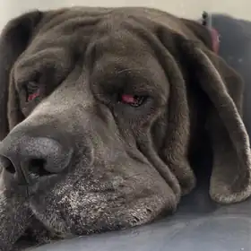
Uno, a Cane Corso with cherry eye in both eyes
Patient
Uno
Species and breed
Dog, Cane Corso
Age
6 years
Came to us with
A red, swollen lump in the corner of both eyes.
What John found
Cherry eye in both eyes: the tear gland in the corner of each eye had slipped out of place.
What we did
Cherry eye surgery on both eyes, using the pocket method. The gland is tucked back into a small pocket of tissue and kept, because it makes tears for the eye.
Recovery
Home with an anti-inflammatory, antibiotics and a cone collar, and a post-op check five days later.
Outcome
Both eyes healing well at his post-op check.
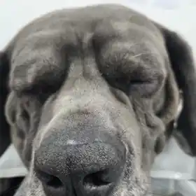
Before surgery, with cherry eye in both eyesFast asleep after his operation